Back Of Neck Anatomy Muscles - Vivian Grisogono About The Back And Neck : Sternohyoid, sternothyroid, thyrohyoid, omohyoid anterior vertebral muscles:
byAdmin•
0
Back Of Neck Anatomy Muscles - Vivian Grisogono About The Back And Neck : Sternohyoid, sternothyroid, thyrohyoid, omohyoid anterior vertebral muscles:. They are divided into three groups, as shown below. These muscles course from your vertebral column to your ribs. The back muscles can be three types. The extrinsic muscles that are associated with upper extremity and shoulder movement, and the intrinsic muscles that deal thick splenius muscles form the superficial layer of muscles and are located on the lateral and posterior portions of the neck. As you know, the neck is the part of the body that sits between the head and torso.
The pll starts at c2 and goes down the back of the vertebral bodies and intervertebral discs. Neck mobility is necessary primarily to rotate the head and keep the head upright. The head rests on the top part of the vertebral column, with the skull joining at c1. Superficial muscles are the muscles closest to the skin surface and can usually be seen while a body is performing actions. The three scalene muscles are found forming the floor of the posterior triangle.
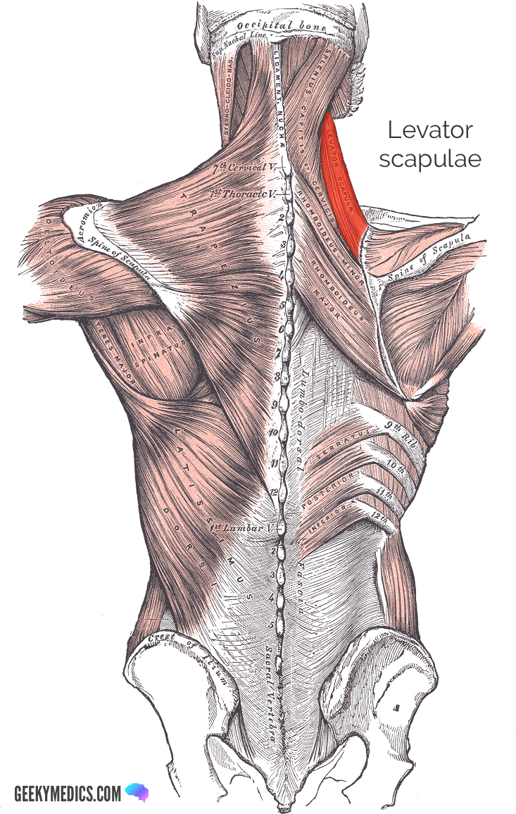
Superficial Back Muscles Anatomy Geeky Medics from geekymedics.com Watch cervical muscle anatomy animation. The extensors and rotators of the head and neck: The suprahyoid muscles originate from above the hyoid bone in the chin region. The three scalene muscles are found forming the floor of the posterior triangle. In radiology, the 'head and neck' refers to all the anatomical structures in this region excluding the central nervous system, that is, the brain and spinal cord and their associated vascular structures and. Superficial muscles are the muscles closest to the skin surface and can usually be seen while a body is performing actions. Integrates anatomy and physiology of cells, tissues, organs, the systems of the human body, and mechanisms responsible for homeostasis. Working in pairs on the left and.
The neck muscles, including the sternocleidomastoid and the trapezius, are responsible for the gross motor movement in the muscular system of the head and neck.
Back muscles are divided into two specific groups: The superficial group acts on upper limbs and. Working in pairs on the left and. There are many muscles around the neck that help to support the cervical spine and allow you to move your head in different directions. The head rests on the top part of the vertebral column, with the skull joining at c1. The posterior muscles of the neck are primarily concerned with head movements, like extension. Muscles of the neck are described separately from the compartments. The posterior muscles of the neck are primarily concerned with head movements, like extension. The back anatomy includes the latissimus dorsi, trapezius, erector spinae, rhomboid, and the teres major. Neck mobility is necessary primarily to rotate the head and keep the head upright. The neck muscles, including the sternocleidomastoid and the trapezius, are responsible for the gross motor movement in the muscular system of the head and neck. There are several individual muscles within the back anatomy, and it's important to take a quick look the image below to shows all the major back muscles (as well as some neck muscles) They are divided into three groups, as shown below.
The head rests on the top part of the vertebral column, with the skull joining at c1. The anatomy of your back muscles can be complex. Digastric, mylohyoid, geniohyoid, stylohyoid infrahyoid muscles: Head and neck anatomy is important when considering pathology affecting the same area. This article covers the anatomy of the deep muscles of the back, including their function, blood supply, innervation, origin and insertion.
Normal Anatomy Of The Deep Muscles Of The Back And Neck Medical Art Works from cdn.shopify.com These muscles course from your vertebral column to your ribs. The posterior muscles of the neck are primarily concerned with head movements, like extension. The extensors and rotators of the head and neck: Many in the neck help to stabilize or move the head. As you know, the neck is the part of the body that sits between the head and torso. The back anatomy includes the latissimus dorsi, trapezius, erector spinae, rhomboid, and the teres major. Back muscles are divided into two specific groups: The anterior and middle scalenes originate from the transverse processes of certain cervical vertebrae and attach to the first rib.
Head and neck anatomy is important when considering pathology affecting the same area. The deep back muscles lie immediately adjacent to the vertebral column and ribs. Neck flexion and homolateral tilt. Extrinsic muscle layers of the back. They work on the hyoid bone, with the suprahyoid muscles pulling up and the infrahyoid. Working in pairs on the left and. The three scalene muscles are found forming the floor of the posterior triangle. There are several individual muscles within the back anatomy, and it's important to take a quick look the image below to shows all the major back muscles (as well as some neck muscles) These muscles course from your vertebral column to your ribs. The neck has no external bone protective structures, so it is quite mobile. The muscles of the anterior neck are arranged to facilitate swallowing and speech. This article covers the anatomy of the deep muscles of the back, including their function, blood supply, innervation, origin and insertion. The anterior and middle scalenes originate from the transverse processes of certain cervical vertebrae and attach to the first rib.
Human muscle anatomy 12 photos of the human muscle anatomy human anatomy muscle questions, human anatomy muscles clay learning system, human muscle anatomy head, human muscle anatomy leg, human muscle. The posterior muscles of the neck are primarily concerned with head movements, like extension. Head and neck anatomy is important when considering pathology affecting the same area. Covers deep muscles of back and trunk. The muscles of the back that work together to support the spine, help keep the body upright and allow twist and bend in many directions.
The Human Muscle System Neck Muscle Anatomy Muscles Of The Neck Sternocleidomastoid Muscle from i.pinimg.com They work on the hyoid bone, with the suprahyoid muscles pulling up and the infrahyoid. Watch cervical muscle anatomy animation. The extensors and rotators of the head and neck: The suprahyoid muscles originate from above the hyoid bone in the chin region. Back muscles are divided into two specific groups: Intermediate back muscles and c. Bones of the neck picture. The pll starts at c2 and goes down the back of the vertebral bodies and intervertebral discs.
Several other muscles of the back also extend up to the neck region and are partly connected with the cervical part of the vertebral column, including the trapezius, levator scapulae, splenius, iliocostalis, longissimus, rotatores, semispinalis, interspinales, and intertransversarii muscles.
Here the extrinsic back muscles are classified into logical subgroups to facilitate knowledge. The neck has no external bone protective structures, so it is quite mobile. The anatomy of your back muscles can be complex. The extensors and rotators of the head and neck: This article covers the anatomy of the deep muscles of the back, including their function, blood supply, innervation, origin and insertion. Extrinsic muscle layers of the back. Covers deep muscles of back and trunk. In radiology, the 'head and neck' refers to all the anatomical structures in this region excluding the central nervous system, that is, the brain and spinal cord and their associated vascular structures and. Sternohyoid, sternothyroid, thyrohyoid, omohyoid anterior vertebral muscles: The muscles of the back and neck that move the vertebral column are complex, overlapping, and can be divided into five groups. Many in the neck help to stabilize or move the head. Muscles of the posterior neck and the back. Superficial muscles are the muscles closest to the skin surface and can usually be seen while a body is performing actions.